
Breakthrough in lung disease diagnostics: first successful use of ultrasound computed tomography (USCT)
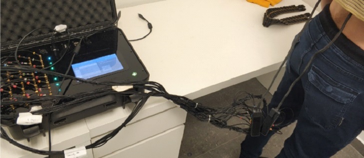
A significant breakthrough has been made in the field of lung diagnostics. Scientists from the Engineering Tomography Laboratory (ETL) at the University of Bath, in collaboration with Netrix S.A., have successfully used ultrasound computed tomography (USCT) to image the lungs of patients for the first time in the world. This pioneering achievement sets a new direction in diagnostic methods, offering a solution that is not only safe and non-invasive, but also portable and available at the patient’s bedside.
Unlike currently used techniques such as computed tomography (CT) or magnetic resonance imaging (MRI), the new method does not require expensive and extensive infrastructure, does not expose the patient to ionizing radiation, and can be used in hospital conditions without the need to transport the patient. Thanks to these features, USCT has the potential to revolutionize patient care, especially in intensive care units, where mobility and speed of response are key.
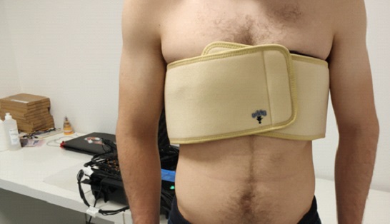
The developed system consists of 32 ultrasonic sensors arranged in a flexible, easy-to-wear chest belt. The device uses low-frequency sound waves (40 kHz) that penetrate lung tissue, recording changes in the speed of wave propagation and the level of their attenuation. This allows for dynamic mapping of changes occurring in the lungs during the respiratory cycle.
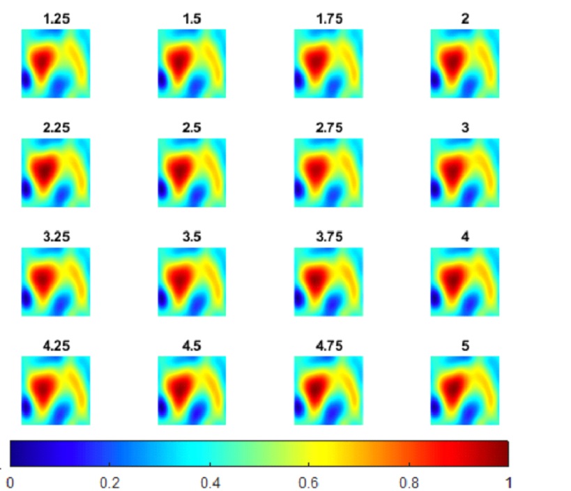
As part of the conducted in vivo studies, the technology was tested on a group of eight healthy volunteers. The results confirmed the ability of USCT to detect changes in the lungs in real time, dependent on the respiratory phase. Advanced machine learning algorithms, such as autoencoders and U-Net neural networks, also played a key role here, enabling precise reconstruction of lung images based on simulation data.
The developed technology demonstrated high sensitivity to changes in lung volume and structure during inhalation and exhalation, which may be of significant importance in the diagnosis and monitoring of the course of many respiratory diseases, including pulmonary edema, pneumothorax, or complications associated with SARS-CoV-2 infection.
The prospects for clinical use of USCT are promising. In the near future, this method may become a standard tool for monitoring mechanical ventilation in ICU patients, support early detection of lung pathologies, and complement other imaging techniques, such as electroimpedance tomography (EIT), to obtain more detailed diagnostic information.
The research results were published in the prestigious IEEE Transactions on Biomedical Engineering journal, and the full text of the publication is available at: link to publication
The research team behind this achievement consists of Prof. Manuchehr Soleimani and Dr. Rinki Goyal from the University of Bath, as well as Prof. Tomasz Rymarczyk from Netrix S.A. Their joint work is a milestone towards the development of accessible, safe and effective medical imaging technologies that can change the way we care for patients with respiratory diseases.